

How Is Lysosome Produced By An Animal Able To Distinguish Between Bacteria And Self Proteins
| Jail cell biology | |
|---|---|
| Animal cell diagram | |
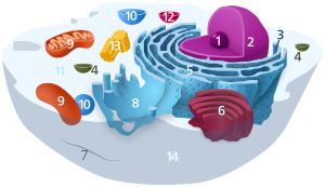 Components of a typical animal prison cell:
|
A lysosome () is a membrane-bound organelle found in many animal cells.[1] They are spherical vesicles that contain hydrolytic enzymes that can pause down many kinds of biomolecules. A lysosome has a specific limerick, of both its membrane proteins, and its lumenal proteins. The lumen's pH (~4.v–v.0)[2] is optimal for the enzymes involved in hydrolysis, analogous to the activity of the stomach. Besides degradation of polymers, the lysosome is involved in various cell processes, including secretion, plasma membrane repair, apoptosis, cell signaling, and energy metabolism.[three]

Lysosomes assimilate materials taken into the cell and recycle intracellular materials. Step ane shows textile entering a food vacuole through the plasma membrane, a procedure known as endocytosis. In footstep two a lysosome with an active hydrolytic enzyme comes into the moving picture equally the food vacuole moves away from the plasma membrane. Step iii consists of the lysosome fusing with the food vacuole and hydrolytic enzymes entering the food vacuole. In the final step, step 4, hydrolytic enzymes digest the food particles.[4]
Lysosomes human action as the waste disposal system of the cell by digesting used materials in the cytoplasm, from both inside and outside the cell. Material from outside the prison cell is taken up through endocytosis, while material from the inside of the cell is digested through autophagy.[v] The sizes of the organelles vary greatly—the larger ones can be more 10 times the size of the smaller ones.[6] They were discovered and named by Belgian biologist Christian de Duve, who somewhen received the Nobel Prize in Physiology or Medicine in 1974.
Lysosomes are known to contain more 60 different enzymes, and have more than fifty membrane proteins.[7] [8] Enzymes of the lysosomes are synthesized in the rough endoplasmic reticulum and exported to the Golgi apparatus upon recruitment by a complex composed of CLN6 and CLN8 proteins.[9] [x] The enzymes are trafficked from the Golgi apparatus to lysosomes in small vesicles, which fuse with larger acidic vesicles. Enzymes destined for a lysosome are specifically tagged with the molecule mannose 6-phosphate, then that they are properly sorted into acidified vesicles.[11] [12]
In 2009, Marco Sardiello and co-workers discovered that the synthesis of almost lysosomal enzymes and membrane proteins is controlled past transcription factor EB (TFEB), which promotes the transcription of nuclear genes.[13] [14] Mutations in the genes for these enzymes are responsible for more than 50 different human genetic disorders, which are collectively known as lysosomal storage diseases. These diseases result from an accumulation of specific substrates, due to the disability to break them downwardly. These genetic defects are related to several neurodegenerative disorders, cancers, cardiovascular diseases, and aging-related diseases.[15] [16] [17]
Discovery [edit]

TEM views of diverse vesicular compartments. Lysosomes are denoted by "Ly". They are dyed dark due to their acidity; in the middle of the top image, a Golgi Appliance tin can be seen, distal from the prison cell membrane relative to the lysosomes.
Christian de Duve, the chairman of the Laboratory of Physiological Chemical science at the Catholic Academy of Louvain in Belgium, had been studying the mechanism of action of a pancreatic hormone insulin in liver cells. By 1949, he and his team had focused on the enzyme called glucose vi-phosphatase, which is the kickoff crucial enzyme in carbohydrate metabolism and the target of insulin. They already suspected that this enzyme played a central role in regulating claret sugar levels. However, even after a series of experiments, they failed to purify and isolate the enzyme from the cellular extracts. Therefore, they tried a more than arduous procedure of cell fractionation, by which cellular components are separated based on their sizes using centrifugation.
They succeeded in detecting the enzyme activity from the microsomal fraction. This was the crucial stride in the serendipitous discovery of lysosomes. To approximate this enzyme activeness, they used that of the standardized enzyme acrid phosphatase and found that the activeness was only 10% of the expected value. One day, the enzyme activity of purified prison cell fractions which had been refrigerated for five days was measured. Surprisingly, the enzyme activity was increased to normal of that of the fresh sample. The result was the same no matter how many times they repeated the estimation, and led to the determination that a membrane-like barrier limited the accessibility of the enzyme to its substrate, and that the enzymes were able to lengthened subsequently a few days (and react with their substrate). They described this membrane-similar barrier as a "saclike construction surrounded by a membrane and containing acrid phosphatase."[18]
It became clear that this enzyme from the jail cell fraction came from membranous fractions, which were definitely cell organelles, and in 1955 De Duve named them "lysosomes" to reverberate their digestive properties.[19] The same year, Alex B. Novikoff from the University of Vermont visited de Duve's laboratory, and successfully obtained the kickoff electron micrographs of the new organelle. Using a staining method for acrid phosphatase, de Duve and Novikoff confirmed the location of the hydrolytic enzymes of lysosomes using light and electron microscopic studies.[twenty] [21] de Duve won the Nobel Prize in Physiology or Medicine in 1974 for this discovery.
Originally, De Duve had termed the organelles the "suicide bags" or "suicide sacs" of the cells, for their hypothesized role in apoptosis.[22] However, it has since been ended that they only play a small role in cell death.[23]
Function and structure [edit]
Lysosomes contain a diversity of enzymes, enabling the prison cell to break down various biomolecules it engulfs, including peptides, nucleic acids, carbohydrates, and lipids (lysosomal lipase). The enzymes responsible for this hydrolysis require an acidic environs for optimal activity.
In addition to being able to break downwards polymers, lysosomes are capable of fusing with other organelles & digesting large structures or cellular droppings; through cooperation with phagosomes, they are able to conduct autophagy, immigration out damaged structures. Similarly, they are able to pause down virus particles or leaner in phagocytosis of macrophages.
The size of lysosomes varies from 0.1 μm to 1.2 μm.[24] With a pH ranging from ~4.5–5.0, the interior of the lysosomes is acidic compared to the slightly basic cytosol (pH 7.2). The lysosomal membrane protects the cytosol, and therefore the rest of the cell, from the degradative enzymes within the lysosome. The cell is additionally protected from any lysosomal acid hydrolases that drain into the cytosol, as these enzymes are pH-sensitive and do non function well or at all in the alkaline environs of the cytosol. This ensures that cytosolic molecules and organelles are non destroyed in case there is leakage of the hydrolytic enzymes from the lysosome.
The lysosome maintains its pH differential past pumping in protons (H+ ions) from the cytosol beyond the membrane via proton pumps and chloride ion channels. Vacuolar-ATPases are responsible for transport of protons, while the counter transport of chloride ions is performed by ClC-7 Cl−/H+ antiporter. In this mode a steady acidic surroundings is maintained.[25] [26]
Information technology sources its versatile chapters for degradation by import of enzymes with specificity for unlike substrates; cathepsins are the major class of hydrolytic enzymes, while lysosomal alpha-glucosidase is responsible for carbohydrates, and lysosomal acrid phosphatase is necessary to release phosphate groups of phospholipids.
Germination [edit]

The lysosome is shown in purple, as an endpoint in endocytotic sorting. AP2 is necessary for vesicle formation, whereas the mannose-half dozen-receptor is necessary for sorting hydrolase into the lysosome'due south lumen.
Many components of animal cells are recycled by transferring them inside or embedded in sections of membrane. For instance, in endocytosis (more specifically, macropinocytosis), a portion of the prison cell's plasma membrane pinches off to form vesicles that will eventually fuse with an organelle within the jail cell. Without active replenishment, the plasma membrane would continuously subtract in size. It is thought that lysosomes participate in this dynamic membrane commutation organisation and are formed by a gradual maturation process from endosomes.[27] [28]
The production of lysosomal proteins suggests i method of lysosome sustainment. Lysosomal poly peptide genes are transcribed in the nucleus in a process that is controlled past transcription factor EB (TFEB).[14] mRNA transcripts leave the nucleus into the cytosol, where they are translated by ribosomes. The nascent peptide bondage are translocated into the crude endoplasmic reticulum, where they are modified. Lysosomal soluble proteins exit the endoplasmic reticulum via COPII-coated vesicles after recruitment by the EGRESS complex (Due eastR-to-One thousandolgi relaying of enzymes of the lysosomal due southystem), which is composed of CLN6 and CLN8 proteins.[ix] [10] COPII vesicles so deliver lysosomal enzymes to the Golgi appliance, where a specific lysosomal tag, mannose 6-phosphate, is added to the peptides. The presence of these tags allow for binding to mannose half-dozen-phosphate receptors in the Golgi apparatus, a miracle that is crucial for proper packaging into vesicles destined for the lysosomal system.[29]
Upon leaving the Golgi appliance, the lysosomal enzyme-filled vesicle fuses with a belatedly endosome, a relatively acidic organelle with an approximate pH of 5.5. This acidic environs causes dissociation of the lysosomal enzymes from the mannose vi-phosphate receptors. The enzymes are packed into vesicles for further transport to established lysosomes.[29] The late endosome itself can somewhen grow into a mature lysosome, as evidenced past the transport of endosomal membrane components from the lysosomes dorsum to the endosomes.[27]
Pathogen entry [edit]
Equally the endpoint of endocytosis, the lysosome also acts as a safeguard in preventing pathogens from being able to reach the cytoplasm before being degraded. Pathogens often hijack endocytotic pathways such every bit pinocytosis in order to gain entry into the cell. The lysosome prevents easy entry into the cell by hydrolyzing the biomolecules of pathogens necessary for their replication strategies; reduced Lysosomal activity results in an increase in viral infectivity, including HIV.[30] In addition, AB5 toxins such as cholera hijack the endosomal pathway while evading lysosomal degradation.[30]
Clinical significance [edit]
Lysosomes are involved in a grouping of genetically inherited deficiencies, or mutations called lysosomal storage diseases (LSD), inborn errors of metabolism acquired by a dysfunction of 1 of the enzymes. The rate of incidence is estimated to be 1 in five,000 births, and the true figure expected to exist higher every bit many cases are likely to be undiagnosed or misdiagnosed. The master crusade is deficiency of an acid hydrolase. Other conditions are due to defects in lysosomal membrane proteins that fail to transport the enzyme, not-enzymatic soluble lysosomal proteins. The initial effect of such disorders is aggregating of specific macromolecules or monomeric compounds inside the endosomal–autophagic–lysosomal organization.[xv] This results in abnormal signaling pathways, calcium homeostasis, lipid biosynthesis and degradation and intracellular trafficking, ultimately leading to pathogenetic disorders. The organs about affected are brain, viscera, os and cartilage.[31] [32]
There is no directly medical treatment to cure LSDs.[33] The most common LSD is Gaucher's disease, which is due to deficiency of the enzyme glucocerebrosidase. Consequently, the enzyme substrate, the fatty acid glucosylceramide accumulates, specially in white blood cells, which in turn affects spleen, liver, kidneys, lungs, brain and bone marrow. The disease is characterized by bruises, fatigue, anaemia, low blood platelets, osteoporosis, and enlargement of the liver and spleen.[34] [35] Equally of 2017, enzyme replacement therapy is bachelor for treating 8 of the l-60 known LDs.[36]
The most astringent and rarely found, lysosomal storage illness is inclusion prison cell disease.[37]
Metachromatic leukodystrophy is another lysosomal storage disease that too affects sphingolipid metabolism.
Dysfunctional lysosome activeness is also heavily implicated in the biology of aging, and historic period-related diseases such equally Alzheimer's, Parkinson's, and cardiovascular disease. [38] [39]
Dissimilar enzymes present in Lysosomes [40] [edit]
| Sr. No | Enzymes | Substrate |
|---|---|---|
| 1 | Phosphates | |
| A- Acrid phosphatase | Most phosphomonoesters | |
| B- Acrid phosphodiesterase | Oligonucleotides and phosphodiesterase | |
| 2 | Nucleases | |
| A- Acid ribonuclease | RNA | |
| B- Acid deoxyribonuclease | DNA | |
| 3 | Polysaccharides/ mucopolysaccharides hydrolyzing enzymes | |
| A- β-Galactosidase | Galactosides | |
| B- α-Glucosidase | Glycogen | |
| C- α-Mannosidase | Mannosides, glycoproteins | |
| D- β- Glucoronidase | Polysaccharides and mucopolysaccharides | |
| E- Lysozymes | Bacterial cell walls and mucopolysaccharides | |
| F- Hyaluronidase | Hyaluronic acids, chondroitin sulfates | |
| H- Arylsulphatase | Organic sulfates | |
| 4 | Proteases | |
| A- Cathepsin(s) | Proteins | |
| B- Collagenase | Collagen | |
| C- Peptidase | Peptides | |
| 5 | Lipid degrading enzymes | |
| A- Esterase | Fatty acyl esters | |
| B- Phospholipase | Phospholipids | |
| 6 | Sulfatases | |
| A- Arylsulfatase(A, B & G) | O- and N-Sulfate esters | |
| B- Glucosamine (N-acetyl)-6-Sulfatase/GNS | Glycosaminoglycans | |
| C- Iduronate ii-Sulfatase/IDS | O- and Due north-Sulfate esters |
Lysosomotropism [edit]
Weak bases with lipophilic backdrop accumulate in acidic intracellular compartments similar lysosomes. While the plasma and lysosomal membranes are permeable for neutral and uncharged species of weak bases, the charged protonated species of weak bases do not permeate biomembranes and accumulate within lysosomes. The concentration inside lysosomes may reach levels 100 to g fold higher than extracellular concentrations. This phenomenon is chosen lysosomotropism,[41] "acid trapping" or "proton pump" effect.[42] The amount of accumulation of lysosomotropic compounds may be estimated using a cell-based mathematical model.[43]
A meaning role of the clinically approved drugs are lipophilic weak bases with lysosomotropic properties. This explains a number of pharmacological backdrop of these drugs, such equally loftier tissue-to-blood concentration gradients or long tissue elimination half-lives; these properties have been constitute for drugs such every bit haloperidol,[44] levomepromazine,[45] and amantadine.[46] Nonetheless, high tissue concentrations and long elimination half-lives are explained also by lipophilicity and absorption of drugs to fatty tissue structures. Important lysosomal enzymes, such equally acid sphingomyelinase, may be inhibited by lysosomally accumulated drugs.[47] [48] Such compounds are termed FIASMAs (functional inhibitor of acid sphingomyelinase)[49] and include for example fluoxetine, sertraline, or amitriptyline.
Ambroxol is a lysosomotropic drug of clinical employ to treat conditions of productive cough for its mucolytic action. Ambroxol triggers the exocytosis of lysosomes via neutralization of lysosomal pH and calcium release from acidic calcium stores.[50] Presumably for this reason, Ambroxol was also institute to amend cellular function in some affliction of lysosomal origin such as Parkinson's or lysosomal storage disease.[51] [52]
Systemic lupus erythematosus [edit]
Impaired lysosome part is prominent in systemic lupus erythematosus preventing macrophages and monocytes from degrading neutrophil extracellular traps[53] and allowed complexes.[54] [55] [56] The failure to dethrone internalized immune complexes stems from chronic mTORC2 activity, which impairs lysosome acidification.[57] As a result, immune complexes in the lysosome recycle to the surface of macrophages causing an accumulation of nuclear antigens upstream of multiple lupus-associated pathologies.[54] [58] [59]
Controversy in botany [edit]
By scientific convention, the term lysosome is applied to these vesicular organelles only in animals, and the term vacuole is applied to those in plants, fungi and algae (some animal cells besides take vacuoles). Discoveries in plant cells since the 1970s started to challenge this definition. Plant vacuoles are constitute to be much more than diverse in structure and function than previously idea.[60] [61] Some vacuoles incorporate their own hydrolytic enzymes and perform the classic lysosomal activity, which is autophagy.[62] [63] [64] These vacuoles are therefore seen as fulfilling the office of the brute lysosome. Based on de Duve'southward description that "only when considered as part of a organization involved directly or indirectly in intracellular digestion does the term lysosome draw a physiological unit", some botanists strongly argued that these vacuoles are lysosomes.[65] However, this is non universally accepted as the vacuoles are strictly non similar to lysosomes, such as in their specific enzymes and lack of phagocytic functions.[66] Vacuoles practise not have catabolic activity and exercise not undergo exocytosis equally lysosomes practice.[67]
Etymology and pronunciation [edit]
The word lysosome (, ) is New Latin that uses the combining forms lyso- (referring to lysis and derived from the Latin lysis, meaning "to loosen", via Ancient Greek λύσις [lúsis]), and -some, from soma, "body", yielding "body that lyses" or "lytic body". The adjectival grade is lysosomal. The forms *lyosome and *lyosomal are much rarer; they use the lyo- class of the prefix but are often treated by readers and editors as mere unthinking replications of typos, which has no doubtfulness been true as oftentimes as non.
Come across also [edit]
- Peroxisome
- Cathelicidin
- Antimicrobial peptides
- Innate immune system
References [edit]
- ^ By convention similar cells in plants are called vacuoles, encounter § Controversy in botany
- ^ Ohkuma S, Poole B (July 1978). "Fluorescence probe measurement of the intralysosomal pH in living cells and the perturbation of pH by various agents". Proceedings of the National Academy of Sciences of the U.s. of America. 75 (7): 3327–31. Bibcode:1978PNAS...75.3327O. doi:10.1073/pnas.75.seven.3327. PMC392768. PMID 28524.
- ^ Settembre C, Fraldi A, Medina DL, Ballabio A (May 2013). "Signals from the lysosome: a command centre for cellular clearance and free energy metabolism". Nature Reviews Molecular Cell Biology. 14 (5): 283–96. doi:x.1038/nrm3565. PMC4387238. PMID 23609508.
- ^ Holtzclaw FW, et al. (2008). AP* Biological science: to Back-trail Biology (eighth AP ed.). Pearson Benjamin Cummings.
- ^ Underwood, Emily (2018). "When the brain's waste disposal system fails". Knowable Magazine. doi:10.1146/knowable-121118-1.
- ^ Lüllmznn-Rauch R (2005). "History and Morphology of Lysosome". In Zaftig P (ed.). Lysosomes (Online-Ausg. 1 ed.). Georgetown, Tex.: Landes Bioscience/Eurekah.com. pp. 1–16. ISBN978-0-387-28957-1.
- ^ Xu H, Ren D (2015). "Lysosomal physiology". Almanac Review of Physiology. 77 (i): 57–lxxx. doi:10.1146/annurev-physiol-021014-071649. PMC4524569. PMID 25668017.
- ^ "Lysosomal Enzymes". world wide web.rndsystems.com. R&D Systems. Retrieved 4 October 2016.
- ^ a b di Ronza A, Bajaj 50, Sharma J, Sanagasetti D, Lotfi P, Adamski CJ, Collette J, Palmieri One thousand, Amawi A, Popp 50, Chang KT, Meschini MC, Leung HE, Segatori L, Simonati A, Sifers RN, Santorelli FM, Sardiello M (December 2018). "CLN8 is an endoplasmic reticulum cargo receptor that regulates lysosome biogenesis". Nature Cell Biological science. 20 (12): 1370–1377. doi:10.1038/s41556-018-0228-vii. PMC6277210. PMID 30397314.
- ^ a b Bajaj L, Sharma J, di Ronza A, Zhang P, Eblimit A, Pal R, Roman D, Collette JR, Berth C, Chang KT, Sifers RN, Jung SY, Weimer JM, Chen R, Schekman RW, Sardiello M (June 2020). "A CLN6-CLN8 complex recruits lysosomal enzymes at the ER for Golgi transfer". J Clin Invest. 130 (8): 4118–4132. doi:10.1172/JCI130955. PMC7410054. PMID 32597833.
- ^ Saftig P, Klumperman J (September 2009). "Lysosome biogenesis and lysosomal membrane proteins: trafficking meets function". Nature Reviews Molecular Cell Biology. 10 (nine): 623–35. doi:x.1038/nrm2745. PMID 19672277. S2CID 24493663.
- ^ Samie MA, Xu H (June 2014). "Lysosomal exocytosis and lipid storage disorders". Journal of Lipid Research. 55 (half-dozen): 995–1009. doi:10.1194/jlr.R046896. PMC4031951. PMID 24668941.
- ^ Underwood, Emily (2018). "When the brain's waste material disposal organization fails". Knowable Magazine. doi:10.1146/knowable-121118-1.
- ^ a b Sardiello M, Palmieri M, di Ronza A, Medina DL, Valenza M, Gennarino VA, Di Malta C, Donaudy F, Embrione V, Polishchuk RS, Banfi South, Parenti G, Cattaneo Eastward, Ballabio A (July 2009). "A gene network regulating lysosomal biogenesis and part". Science. 325 (5939): 473–7. Bibcode:2009Sci...325..473S. doi:10.1126/scientific discipline.1174447. PMID 19556463. S2CID 20353685.
- ^ a b Platt FM, Boland B, van der Spoel AC (November 2012). "The cell biology of affliction: lysosomal storage disorders: the cellular impact of lysosomal dysfunction". The Journal of Prison cell Biology. 199 (5): 723–34. doi:10.1083/jcb.201208152. PMC3514785. PMID 23185029.
- ^ He LQ, Lu JH, Yue ZY (May 2013). "Autophagy in ageing and ageing-associated diseases". Acta Pharmacologica Sinica. 34 (5): 605–11. doi:10.1038/aps.2012.188. PMC3647216. PMID 23416930.
- ^ Carmona-Gutierrez, Didac; Hughes, Adam L.; Madeo, Frank; Ruckenstuhl, Christoph (1 December 2016). "The crucial touch of lysosomes in aging and longevity". Ageing Research Reviews. Lysosomes in Aging. 32: 2–12. doi:10.1016/j.arr.2016.04.009. ISSN 1568-1637. PMC5081277. PMID 27125853.
- ^ Susana Castro-Obregon (2010). "The Discovery of Lysosomes and Autophagy". Nature Pedagogy. 3 (nine): 49.
- ^ de Duve C (September 2005). "The lysosome turns fifty". Nature Cell Biology. vii (9): 847–9. doi:x.1038/ncb0905-847. PMID 16136179. S2CID 30307451.
- ^ Novikoff AB, Beaufay H, De Duve C (July 1956). "Electron microscopy of lysosomerich fractions from rat liver". The Journal of Biophysical and Biochemical Cytology. two (iv Suppl): 179–84. doi:10.1083/jcb.2.4.179. PMC2229688. PMID 13357540.
- ^ Klionsky DJ (August 2008). "Autophagy revisited: a conversation with Christian de Duve". Autophagy. iv (6): 740–three. doi:10.4161/auto.6398. PMID 18567941.
- ^ Hayashi, Teru, and others. "Subcellular Particles." Subcellular Particles., 1959.
- ^ Turk B, Turk V (2009). "Lysosomes as 'Suicide Numberless' in Prison cell Death: Myth or Reality?". The Journal of Biological Chemistry. 284 (33): 21783–87. doi:x.1074/jbc.R109.023820. PMC2755904. PMID 19473965.
- ^ Kuehnel Due west (2003). Color Atlas of Cytology, Histology, & Microscopic Anatomy (4th ed.). Thieme. p. 34. ISBN978-1-58890-175-0.
- ^ Mindell JA (2012). "Lysosomal acidification mechanisms". Annual Review of Physiology. 74 (1): 69–86. doi:x.1146/annurev-physiol-012110-142317. PMID 22335796.
- ^ Ishida Y, Nayak S, Mindell JA, Grabe M (June 2013). "A model of lysosomal pH regulation". The Journal of Full general Physiology. 141 (6): 705–twenty. doi:10.1085/jgp.201210930. PMC3664703. PMID 23712550.
- ^ a b Alberts B, et al. (2002). Molecular biology of the cell (fourth ed.). New York: Garland Science. ISBN978-0-8153-3218-3.
- ^ Falcone S, Cocucci E, Podini P, Kirchhausen T, Clementi Eastward, Meldolesi J (November 2006). "Macropinocytosis: regulated coordination of endocytic and exocytic membrane traffic events". Journal of Cell Science. 119 (Pt 22): 4758–69. doi:10.1242/jcs.03238. PMID 17077125.
- ^ a b Lodish H, et al. (2000). Molecular prison cell biology (fourth ed.). New York: Scientific American Books. ISBN978-0-7167-3136-eight.
- ^ a b Wei BL, Denton Prisoner of war, O'Neill E, Luo T, Foster JL, Garcia JV (May 2005). "Inhibition of lysosome and proteasome function enhances man immunodeficiency virus type 1 infection". Journal of Virology. 79 (9): 5705–12. doi:x.1128/jvi.79.9.5705-5712.2005. PMC1082736. PMID 15827185.
- ^ Schultz ML, Tecedor L, Chang Thou, Davidson BL (August 2011). "Clarifying lysosomal storage diseases". Trends in Neurosciences. 34 (eight): 401–10. doi:10.1016/j.tins.2011.05.006. PMC3153126. PMID 21723623.
- ^ Lieberman AP, Puertollano R, Raben N, Slaugenhaupt S, Walkley SU, Ballabio A (May 2012). "Autophagy in lysosomal storage disorders". Autophagy. viii (5): 719–30. doi:ten.4161/auto.19469. PMC3378416. PMID 22647656.
- ^ .Parenti G, Pignata C, Vajro P, Salerno M (January 2013). "New strategies for the treatment of lysosomal storage diseases (review)". International Periodical of Molecular Medicine. 31 (1): 11–xx. doi:10.3892/ijmm.2012.1187. PMID 23165354.
- ^ Rosenbloom BE, Weinreb NJ (2013). "Gaucher disease: a comprehensive review". Critical Reviews in Oncogenesis. xviii (iii): 163–75. doi:10.1615/CritRevOncog.2013006060. PMID 23510062.
- ^ Sidransky E (October 2012). "Gaucher illness: insights from a rare Mendelian disorder". Discovery Medicine. 14 (77): 273–81. PMC4141347. PMID 23114583.
- ^ Solomon Chiliad, Muro S (September 2017). "Lysosomal enzyme replacement therapies: Historical evolution, clinical outcomes, and future perspectives". Advanced Drug Commitment Reviews. 118: 109–134. doi:x.1016/j.addr.2017.05.004. PMC5828774. PMID 28502768.
- ^ Alberts, Bruce (2002). Molecular biological science of the cell (quaternary ed.). Garland Science. p. 744. ISBN978-0815340720.
- ^ Carmona-Gutierrez, Didac; Hughes, Adam 50.; Madeo, Frank; Ruckenstuhl, Christoph (1 December 2016). "The crucial impact of lysosomes in crumbling and longevity". Ageing Research Reviews. Lysosomes in Crumbling. 32: 2–12. doi:10.1016/j.arr.2016.04.009. ISSN 1568-1637. PMC5081277. PMID 27125853.
- ^ Finkbeiner, Steven (1 Apr 2019). "The Autophagy Lysosomal Pathway and Neurodegeneration". Cold Spring Harbor Perspectives in Biology. 12 (3): a033993. doi:10.1101/cshperspect.a033993. ISSN 1943-0264. PMC6773515. PMID 30936119.
- ^ Pranav Kumar. (2013). Life Sciences : Fundamentals and do. Mina, Usha. (third ed.). New Delhi: Pathfinder Academy. ISBN978-81-906427-0-five. OCLC 857764171.
- ^ de Duve C, de Barsy T, Poole B, Trouet A, Tulkens P, Van Hoof F (September 1974). "Commentary. Lysosomotropic agents". Biochemical Pharmacology. 23 (18): 2495–531. doi:x.1016/0006-2952(74)90174-9. PMID 4606365.
- ^ Traganos F, Darzynkiewicz Z (1994). "Lysosomal proton pump activity: supravital jail cell staining with acridine orangish differentiates leukocyte subpopulations". Methods Cell Biol. 41: 185–94. doi:10.1016/S0091-679X(08)61717-iii. PMID 7532261.
- ^ Trapp S, Rosania GR, Horobin RW, Kornhuber J (October 2008). "Quantitative modeling of selective lysosomal targeting for drug blueprint". European Biophysics Journal. 37 (8): 1317–28. doi:10.1007/s00249-008-0338-4. PMC2711917. PMID 18504571.
- ^ Kornhuber J, Schultz A, Wiltfang J, Meineke I, Gleiter CH, Zöchling R, Boissl KW, Leblhuber F, Riederer P (June 1999). "Persistence of haloperidol in man encephalon tissue". The American Periodical of Psychiatry. 156 (6): 885–90. doi:10.1176/ajp.156.half-dozen.885. PMID 10360127. S2CID 7258546.
- ^ Kornhuber J, Weigmann H, Röhrich J, Wiltfang J, Bleich Due south, Meineke I, Zöchling R, Härtter Southward, Riederer P, Hiemke C (March 2006). "Region specific distribution of levomepromazine in the human encephalon". Journal of Neural Manual. 113 (3): 387–97. doi:10.1007/s00702-005-0331-3. PMID 15997416. S2CID 24735371.
- ^ Kornhuber J, Quack Chiliad, Danysz W, Jellinger K, Danielczyk Westward, Gsell Due west, Riederer P (July 1995). "Therapeutic brain concentration of the NMDA receptor antagonist amantadine". Neuropharmacology. 34 (seven): 713–21. doi:10.1016/0028-3908(95)00056-c. PMID 8532138. S2CID 25784783.
- ^ Kornhuber J, Tripal P, Reichel Thou, Terfloth L, Bleich South, Wiltfang J, Gulbins E (January 2008). "Identification of new functional inhibitors of acid sphingomyelinase using a structure-property-activity relation model". Journal of Medicinal Chemistry. 51 (2): 219–37. CiteSeerX10.i.1.324.8854. doi:ten.1021/jm070524a. PMID 18027916.
- ^ Kornhuber J, Muehlbacher M, Trapp S, Pechmann S, Friedl A, Reichel Grand, Mühle C, Terfloth L, Groemer TW, Spitzer GM, Liedl KR, Gulbins E, Tripal P (2011). Riezman H (ed.). "Identification of novel functional inhibitors of acid sphingomyelinase". PLOS ONE. 6 (eight): e23852. Bibcode:2011PLoSO...623852K. doi:10.1371/periodical.pone.0023852. PMC3166082. PMID 21909365.
- ^ Kornhuber J, Tripal P, Reichel One thousand, Mühle C, Rhein C, Muehlbacher M, Groemer TW, Gulbins Due east (2010). "Functional Inhibitors of Acid Sphingomyelinase (FIASMAs): a novel pharmacological grouping of drugs with wide clinical applications". Cellular Physiology and Biochemistry. 26 (one): 9–20. doi:10.1159/000315101. PMID 20502000.
- ^ Fois G, Hobi North, Felder Eastward, Ziegler A, Miklavc P, Walther P, Radermacher P, Haller T, Dietl P (December 2015). "A new part for an former drug: Ambroxol triggers lysosomal exocytosis via pH-dependent Ca²⁺ release from acidic Ca²⁺ stores". Cell Calcium. 58 (vi): 628–37. doi:10.1016/j.ceca.2015.x.002. PMID 26560688.
- ^ Albin RL, Dauer WT (May 2014). "Magic shotgun for Parkinson'due south affliction?". Brain. 137 (Pt 5): 1274–v. doi:x.1093/brain/awu076. PMID 24771397.
- ^ McNeill A, Magalhaes J, Shen C, Chau KY, Hughes D, Mehta A, Foltynie T, Cooper JM, Abramov AY, Gegg One thousand, Schapira AH (May 2014). "Ambroxol improves lysosomal biochemistry in glucocerebrosidase mutation-linked Parkinson disease cells". Brain. 137 (Pt 5): 1481–95. doi:10.1093/brain/awu020. PMC3999713. PMID 24574503.
- ^ Hakkim A, Fürnrohr BG, Amann Thousand, Laube B, Abed UA, Brinkmann V, Herrmann M, Voll RE, Zychlinsky A (May 2010). "Impairment of neutrophil extracellular trap degradation is associated with lupus nephritis". Proceedings of the National Academy of Sciences of the Usa of America. 107 (21): 9813–viii. Bibcode:2010PNAS..107.9813H. doi:10.1073/pnas.0909927107. PMC2906830. PMID 20439745.
- ^ a b Monteith AJ, Kang S, Scott E, Hillman Thou, Rajfur Z, Jacobson K, Costello MJ, Vilen BJ (Apr 2016). "Defects in lysosomal maturation facilitate the activation of innate sensors in systemic lupus erythematosus". Proceedings of the National Academy of Sciences of the U.s.a. of America. 113 (xv): E2142–51. Bibcode:2016PNAS..113E2142M. doi:10.1073/pnas.1513943113. PMC4839468. PMID 27035940.
- ^ Kavai Chiliad, Szegedi G (August 2007). "Immune circuitous clearance past monocytes and macrophages in systemic lupus erythematosus". Autoimmunity Reviews. 6 (vii): 497–502. doi:x.1016/j.autrev.2007.01.017. PMID 17643939.
- ^ Kávai M, Csipö I, Sonkoly I, Csongor J, Szegedi GY (November 1986). "Defective immune circuitous degradation past monocytes in patients with systemic lupus erythematosus". Scandinavian Journal of Immunology. 24 (v): 527–32. doi:x.1111/j.1365-3083.1986.tb02167.x. PMID 3787186. S2CID 23685272.
- ^ Monteith AJ, Vincent HA, Kang S, Li P, Claiborne TM, Rajfur Z, Jacobson One thousand, Moorman NJ, Vilen BJ (July 2018). "mTORC2 Activeness Disrupts Lysosome Acidification in Systemic Lupus Erythematosus by Impairing Caspase-1 Cleavage of Rab39a". Journal of Immunology. 201 (2): 371–382. doi:ten.4049/jimmunol.1701712. PMC6039264. PMID 29866702.
- ^ Kang S, Rogers JL, Monteith AJ, Jiang C, Schmitz J, Clarke SH, Tarrant TK, Truong YK, Diaz M, Fedoriw Y, Vilen BJ (May 2016). "Apoptotic Debris Accumulates on Hematopoietic Cells and Promotes Illness in Murine and Human Systemic Lupus Erythematosus". Journal of Immunology. 196 (ten): 4030–9. doi:ten.4049/jimmunol.1500418. PMC4868781. PMID 27059595.
- ^ Kang S, Fedoriw Y, Brenneman EK, Truong YK, Kikly Chiliad, Vilen BJ (April 2017). "BAFF Induces Third Lymphoid Structures and Positions T Cells within the Glomeruli during Lupus Nephritis". Journal of Immunology. 198 (7): 2602–2611. doi:10.4049/jimmunol.1600281. PMC5360485. PMID 28235864.
- ^ Marty F (April 1999). "Establish vacuoles". The Plant Cell. 11 (4): 587–600. doi:x.2307/3870886. JSTOR 3870886. PMC144210. PMID 10213780.
- ^ Samaj J, Read ND, Volkmann D, Menzel D, Baluska F (August 2005). "The endocytic network in plants". Trends in Cell Biology. xv (8): 425–33. doi:10.1016/j.tcb.2005.06.006. PMID 16006126.
- ^ Matile, P (1978). "Biochemistry and Role of Vacuoles". Almanac Review of Establish Physiology. 29 (1): 193–213. doi:10.1146/annurev.pp.29.060178.001205.
- ^ Moriyasu Y, Ohsumi Y (August 1996). "Autophagy in Tobacco Intermission-Cultured Cells in Response to Sucrose Starvation". Establish Physiology. 111 (iv): 1233–1241. doi:x.1104/pp.111.4.1233. PMC161001. PMID 12226358.
- ^ Jiao BB, Wang JJ, Zhu XD, Zeng LJ, Li Q, He ZH (January 2012). "A novel protein RLS1 with NB-ARM domains is involved in chloroplast degradation during leaf senescence in rice". Molecular Plant. 5 (1): 205–17. doi:10.1093/mp/ssr081. PMID 21980143.
- ^ Swanson SJ, Bethke PC, Jones RL (May 1998). "Barley aleurone cells contain two types of vacuoles. Label Of lytic organelles by use of fluorescent probes". The Plant Cell. x (five): 685–98. doi:10.2307/3870657. JSTOR 3870657. PMC144374. PMID 9596630.
- ^ Holtzman E (1989). Lysosomes. New York: Plenum Press. pp. 7, fifteen. ISBN978-0306-4-3126-five.
- ^ De DN (2000). Institute Cell Vacuoles: An Introduction. Australia: Csiro Publishing. ISBN978-0-643-09944-9.
External links [edit]
| | Look up lysosome in Wiktionary, the free dictionary. |
-
 This article incorporates public domain material from the NCBI certificate: "Scientific discipline Primer".
 - 3D structures of proteins associated with lysosome membrane
- Hide and Seek Foundation For Lysosomal Research
- Lysosomal Disease Network, a inquiry consortium funded by the NIH through its NCATS/Rare Diseases Clinical Research Network
- Self-Destructive Behavior in Cells May Hold Key to a Longer Life
- Mutations in the Lysosomal Enzyme–Targeting Pathway and Persistent Stuttering
- Animation showing how lysosomes are made, and their function
Source: https://en.wikipedia.org/wiki/Lysosome
Posted by: stephenbuslow.blogspot.com
0 Response to "How Is Lysosome Produced By An Animal Able To Distinguish Between Bacteria And Self Proteins"
Post a Comment